
Liver Anatomy
The anatomy of the liver must be understood to truly understand the functioning of this vital organ. Of course this is required for all organs but, as usual, I digress...
🪅
The liver sits in the upper-abdominal region of most mammalian organisms situated beneath the diaphragm. The diaphragm is the muscle that separates the thoracic (chest) cavity from the abdominal (belly) cavity as well as assisting an organism with breathing.
❓
Thought provoking question (based on the above paragraph) - what might happen in a patient whose liver is severely enlarged? I saw this in my clinical rotations and this patient stumped the docs for a brief time as this clinical sign/symptom is not immediately thought of as being caused from liver enlargement.
🐳
Check out the image of the liver, gallbladder, stomach, spleen, pancreas, and small intestines of a human with its associated blood vessels/biliary ducts:
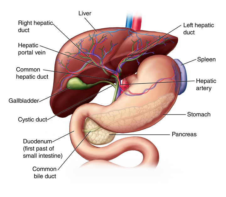
Photo from: https://www.hopkinsmedicine.org/-/media/images/health/1_-conditions/liver-gallbladder-and-pancreas/liver-anatomy.png
Liver Lobes
Liver lobes are macroscopic (visible with the naked eye) sections of the liver that can be seen when looking at a liver. The number and names of the liver lobes is dependent on species. Some examples are below:
Dogs - 6 lobes
Cats- 6 lobes
Horses - 5 lobes
Cattle - 4 lobes
Whales - 2 lobes
Pigs - 5 lobes
Humans - 4 lobes
Image of a Pig Liver with 5 Liver Lobes:

Photo from: https://www.researchgate.net/figure/Anatomic-features-of-pig-liver-a-Pig-liver-showing-relative-position-of-each-of-the_fig4_23803968
Liver Lobules
The liver is often discussed in microscopic (cannot be seen with the naked eye) terms as liver lobules. A liver lobule is a small grouping of liver tissues that make up the functional unit of the liver. A functional unit of an organ is the smallest categorization of an organ that can perform all of the organ's function. The liver lobule is the smallest categorization of the liver that can perform every basic function the liver needs to keep an organism healthy.
💾
There are three (3) major parts of a liver lobule:
1. Portal Triad (five (5) parts)
1. Proper hepatic artery (portal arteriole) - Supplies oxygen to the liver, this vessel is an arteriole off the hepatic artery
2. Hepatic portal vein (portal venule) - High in nutrients and low in oxygen, this vessel is a venule off the portal vein
3. Bile ductules (bile duct) - Branches of the biliary system, these ducts move bile through the liver
4. Lymphatic vessels - Vessels which move lymph through the liver (not pictured in image below)
5. Branch of the vagus nerve - Nerve innervation from the vagus nerve, autonomic nervous system (not pictured in image below)
2. Central vein - Venule that drains deoxygenated hepatic blood into the hepatic vein
3. Hepatocytes - Liver cells

Photo from: https://upload.wikimedia.org/wikipedia/commons/b/b7/2423_Microscopic_Anatomy_of_Liver.jpg
Portal Vein Anatomy
The portal vein is arguably the most important vessel to understand within the liver because it collects all of the blood that comes from the gastrointestinal system (large intestines, small intestines, stomach, etc.), gallbladder, pancreas, and spleen. Now, think about what the previous statement means - what does the portal vein carry?
🍛
You guessed it - the portal vein carries EVERYTHING the gastrointestinal tract absorbs and brings it to the liver. This means the portal vein carries a HUGE amount of nutrients from food, water, microorganisms, toxins, bile salts, etc. The large amount of microscopic materials present in the portal vein explains the major functions of the liver due to its need to process all of the different contents of the portal vein upon its arrival to the liver.
🌟
*In a normal, healthy organism, the portal vein will arrive to the liver and the blood will be processed by the liver PRIOR TO returning to the right side of the heart.* (thought provoking question - why is this important?)
🛼
Below is an image that illustrates how all the blood from the gastrointestinal tract flows into the portal vein. This blood then arrives to and is processed by the liver prior to returning to the heart.

Photo from: https://projects.cos.ncsu.edu/bio370/wrap/TopicsFolder/circulatory3/text_images/portal_vein.jpg
Now that we understand the basic anatomy of the liver, we will continue to explore the different functions of the liver to allow us to work through some upcoming liver-related cases. Keep up the great work and catch you all in the next post (which will hopefully happen in a quicker time frame)!

