Tuesday, October 31, 2023

Test Case Tuesday: Elaine Emits Exhaustion (Case 3)

Picture of stuffed Alpaca named Professor Pocky wearing a stethoscope 

Picture of Professor Pocky taken by Dr. Haus

Click For Explanation of Case Flow


Signalment:

Elaine is a 65-year-old female intact elephant living in an elephant sanctuary in Thailand

🐘

History:

Elaine has been living at the sanctuary her whole life because her mother was killed by poachers when she was a calf.  Elaine’s only pertinent medical history is that she had a suspected viral infection that cleared about ten years ago with supportive treatment.  

Over the past few months, it has been noted that Elaine is appearing more lethargic (tired) and has a pot-bellied appearance.  Today, Elaine collapsed in her habitat and the veterinarian was called in to examine Elaine.

None of the other elephants in the habitat are showing any signs of disease.​

πŸš‘ 

Physical Exam Findings:


Neck:

Jugular vein distension and edema noted in ventral neck 🧨


Cardiovascular:

Increased heart rate, arrhythmia noted, weakened pulses πŸ«€


Abdominal:

Ascites noted (fluid in the abdomen) πŸ«„πŸ½


🩺


πŸ›‘STOP and brainstorm what diagnostic tests you would like to perform on Elaine πŸ›‘


Caution Sign Saying What Diagnostic Testing Should Be Performed?


Diagnostic Testing Results:


Radiographs (X-Rays):

Enlarged heart noted, enlarged vessels leading into the right atrium


Echocardiogram:

All four heart chambers dilated with the right atrium being the most dilated.  No abnormalities of the valves, decreased cardiac output


Electrocardiogram:

Atrial arrhythmia noted 

πŸ§ͺ

Day 1 Conclusion

Before Thoughtful Thursday think about your...

πŸ“‹ Differentials List

πŸ§ͺ Further Diagnostic Testing

πŸ“š Review Material Related to Elaine's Case (i.e. specific diseases of interest, arrhythmias, causes of ascites, heart anatomy, types of heart disease, personal interests, confusing points, etc.)


Thoughtful Thursday:  Elaine Emits Exhaustion (link will go live Thursday, 11/02/2023)

Dr. Haus's Unsolicited Life Advice:  Keep Trying

Heart Anatomy


Photo of Nathan taken by Dr. Haus

The Heart

The heart is the organ that pumps the blood throughout the entire body.  The heart plus all the blood vessels of the body is called the cardiovascular system.  The cardiovascular system ensures that nutrients are brought to and all waste materials are brought away from the cells of the body.

The cardiovascular system is essentially a simple plumbing system.  There are a large amount of pipes of various sizes (blood vessels) and one pump (the heart) that keeps the water (blood) moving.  Understanding the anatomy of the heart is critical to understanding how the heart functions and what is occurring in a sick heart.

πŸ’–

Heart Anatomy

The heart consists of three types of structures:  blood vessels, heart chambers, and heart valves.  Each component of the heart allows the heart to achieve its function of moving blood throughout the body.

πŸ«€

Blood Vessels

The major blood vessels of the heart are the cranial (superior) vena cava, the caudal (inferior) vena cava, the pulmonary artery, the pulmonary vein, and the aorta.  The terms cranial and caudal vena cava are the term for these vessels in quadruped species (species that walk on four legs).  The terms superior and inferior are used in bipedal species such as humans who walk on two legs.

The cranial (superior) vena cava, caudal (inferior) vena cava, and pulmonary artery all carry deoxygenated blood which is blood that has a LOWER amount of oxygen in it.  This blood needs to be delivered to the lungs where carbon dioxide is removed from the blood while oxygen is placed into the blood.  This is done through the process of breathing.  

Remember, an artery moves blood AWAY from the heart regardless of whether the vessel contains a high or low amount of oxygen in it.

The pulmonary vein and aorta carry oxygenated blood which is blood that has a HIGHER amount of oxygen in it.  Oxygenated blood needs to be delivered to the cells of the body to bring them oxygen.  This is done through the heart beating and the blood moving within the vascular system.

Remember, a vein carries blood TOWARDS the heart regardless of whether the vessel contains a high or low amount of oxygen in it.

🧨

Heart Chambers

There are four (4) heart chambers in mammals, birds, and crocodilians.  These heart chambers collect blood while the heart is relaxing and then push the blood in a forwards direction when the heart is beating.  The four (4) heart chambers are the right atrium, the left atrium, the right ventricle, and the left ventricle.

⛰️

Heart Valves

Lastly, the four-chambered heart contains four (4) heart valves.  The heart valves prevent the blood from flowing in a backwards direction.  Remember, the blood must move in the forwards direction or else the blood will not be able to flow throughout the body.  When blood moves backwards in a disease state, the body will become sick as there will not be enough oxygen getting to the cells of the body.

The four (4) heart valves are the tricuspid (right atrioventricular) valve, the bicuspid (mitral or left atrioventricular valve), the pulmonary valve, and the aortic valve.

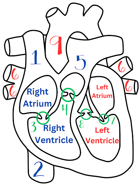
Image of the heart with the heart anatomy labeled

Key:

1.  Cranial (superior) vena cava

2.  Caudal (inferior) vena cava

3.  Tricuspid (right atrioventricular) valve

4.  Pulmonary valve

5.  Pulmonary artery

6.  Pulmonary veins

7.  Bicuspid/mitral (left atrioventricular) valve

8.  Aortic valve

9.  Aorta

πŸ₯ 


Dr. Haus's Unsolicited Life Advice:  Learn the anatomy

What is Cardiac Output and Why is it Important?

Picture of water and a tree line

Photo by Dr. Haus

Cardiac Output

Cardiac output, called CO for short, is the amount of blood that flows out of the left ventricle per minute.  The easier way to define cardiac output is the amount of blood that leaves the heart each time the heart beats.

 πŸ©ΈπŸ«€

Why is Cardiac Output Important?

Cardiac output is extremely important as it is measuring the amount of blood that is being pump throughout the body.  If the cardiac output is too low, that means not enough blood is being pumped out of the heart.  When this occurs, the cells of the body are not receiving the oxygen they need to survive.  The body works EXCEPTIONALLY hard to maintain the correct cardiac output to keep the body alive and functioning. 


Dr. Haus's Unsolicited Life Advice:  Make time for movement daily


What is an Electrocardiogram and Why is it Important?

Picture of a cat named Melvin

Photo by Dr. Haus

Electrocardiogram

An electrocardiogram, called an ECG/EKG for short, is a way to measure the electrical signals in the heart.

 πŸ”ŒπŸ«€

Why is an Electrocardiogram Important?

The beating of a heart relies on electrical signals that tell the heart to beat in the correct manner.  The ECG allows medical professionals to measure the electrical signals in the heart to determine if there is an electrical problem that needs to be addressed in the patient.


Dr. Haus's Unsolicited Life Advice:  It’s not working? Unplug it and plug it back in

What is an Echocardiogram and Why is it Important?

Two cats laying on a bed

Photo by Dr. Haus

Echocardiogram

An echocardiogram, also called an echo for short, is an ultrasound of the heart.  An echocardiogram shows all of the anatomy of the heart and its functioning in real time. 

πŸ«€


Why is an Echocardiogram Important?

An echocardiogram is important because it gives medical professionals a way to see the heart to determine if there are any changes in the heart that require treatment.  The echocardiogram can be thought of as the best way to visualize the heart in real-time to determine if there are any changes.


Dr. Haus's Unsolicited Life Advice:  Take the time to learn how to perform an ultrasound

What are Radiographs (X-rays) and Why are they Important?

Picture of a cat on a couch

Photo by Dr. Haus

Radiographs

A radiograph, also called an x-ray, is a way to visualize the inside of the body at a specific point in time.  Radiographs are taken by a machine and are essentially taking a picture of the inside of the patient's body.

🩻


Why is a Radiograph Important?

A radiograph is important because it gives medical professionals a way to see the inside of the body to determine if there are any obvious causes of disease.  Radiographs are good at diagnosing bone diseases and certain diseases of the internal organs like the stomach, intestines, or lungs.


Dr. Haus's Unsolicited Life Advice:  Always get at least two radiographic views

What is an Arrhythmia and Why is it Important?

Picture of turtles on a rock

Photo by Dr. Haus

Arrhythmia

An arrhythmia is an abnormal heart rhythm.  You can think of this as a time when a heart is not beating in its normal pattern.

πŸ«€


Why is an Arrhythmia Important?

Arrhythmias are important because a heart that is beating out of pattern is a heart that cannot pump blood effectively to the body.  When a patient has an arrhythmia, it is important to diagnose and treat the condition as quickly as possible.


Dr. Haus's Unsolicited Life Advice:  Walk to the beat of your own drum

What is Edema and Why is it Important?

A cat sitting on a couch arm

Photo by Dr. Haus

Edema

Edema is swelling that happens when there is too much water pooling in an area of the body.  Scientifically, edema is when there is more than the normal amount of water in the interstitial fluid space (ISF) and the intracellular fluid space (ICF).

πŸ₯Ό


Why is Edema Important?

Edema is important because it is a sign of injury to a specific area of the body.  Edema is an indication that water balance is not normal and it is a sign to a medical professional that an injury has occurred in that specific location.


Dr. Haus's Unsolicited Life Advice:  Ice, ice, ice an injury

What is the Jugular Vein and Why is it Important?

Picture of a beach

Photo by Dr. Haus

Jugular Vein

The jugular vein is the major vein that brings the deoxygenated blood from the head back to the right side of the heart.  There is a left jugular vein and a right jugular vein.

πŸ”·


Why is the Jugular Vein Important?

The jugular vein is important because it is the way the blood from the head gets back to the heart and lungs to get more oxygen and to get rid of the carbon dioxide.  Given the jugular veins' location in the neck, it can be seen relatively easily and is a location where blood can be drawn or fluids given in a patient.


Dr. Haus's Unsolicited Life Advice:  Jugular sticks seem scary but are great!

Saturday, October 28, 2023

Conclusion Caturday: Florence Feels Feeble (Case 2)


Picture of stuffed alpaca Professor Pocky and stuffed camel Douglas
Photo of TA Douglas (Left) and Professor Pocky (Right) taken by Dr. Haus
 

Click For Explanation of Case Flow


Click for Test Case Tuesday:  Florence Feels Feeble


Click for Thoughtful Thursday:  Florence Feels Feeble 


Quick Review


Diagnosis:

🌌CHRONIC KIDNEY DISEASE (FAILURE)🌌


Pathophysiological Point:

Florence is suffering from a disease where her body's nephrons (kidney cells) are slowly dyingThis means her kidneys are slowly shutting down.


 Questions, Answers, and Further Information:


Level 1 Questions, Answers, and Further Information:

  1. Explain what the kidneys do and why they are important.

    The major kidney function are:  filtering the blood of waste products, regulating blood pressure, regulate production of red blood cells, breakdown of drugs, metabolism of hormones, synthesize vitamin D, manage electrolyte levels, and maintaining blood pH. 

    All the above functions of the kidney affect EVERY other organ system in the body.  The body cannot function very long if the kidneys are not able to complete all of their functions accurately.


  2. If Florence has dilute urine, what does she need to have access to AT ALL TIMES?  (HINT:  Dilute urine means Florence is peeing out more water than normal)

    Florence needs 24/7 access to WATER.  Her dilute urine means Florence is losing a lot of water in her urine and she will need to drink more water to replace all the water she is losing.  Florence is losing water because one of the kidneys jobs is to keep water in the body but if her kidneys are not working the water is lost in the urine instead of kept in the body.

  3. Challenge question, why does Florence have a low number of red blood cells in her body?  (HINT:  For this one you will need to do some information gathering on the kidneys' different jobs)

    Florence has a low number of red blood cells in her body because one of the kidneys' jobs is to tell the body to make more red blood cells.  If the kidneys are slowly failing, they cannot release a hormone called erythropoietin (EPO).  Without erythropoietin, the body does not know it needs to produce and release more red blood cells.

    Helpful Links:


Level 2 Questions, Answers, and Further Information:

  1. Explain pathophysiologically why Florence has pale gums on physical exam and is anemic on blood work.  Be sure to explain all the steps of the process that is NOT occurring in Florence due to her chronic kidney disease.

    Florence has pale gums due to her anemia. This anemia is caused by Florence's Chronic Kidney disease and the dying of her nephrons. The nephrons are responsible for releasing erythropoietin (EPO) into the bloodstream. After the kidneys release EPO, it travels to the bone marrow to stimulate erythropoiesis. This causes the bone marrow to produce and release red blood cells. With Chronic Kidney Disease, the kidneys can no longer produce enough EPO and the bone marrow is not receiving the stimulation to produce enough red blood cells for the body.

  2. Urea is a waste product that is normally urinated out of a patient's body via their kidneys and is an acid.  What acid-base status would you expect in Florence's bloodstream?

    Florence's acid-base status would be a metabolic acidosis (must have both terms to be correct).

  3. Challenge question, what TWO compensatory mechanisms are activated in Florence's body (there are two – assume the juxtaglomerular cells are still functional)?  (HINT:  Think about Florence's dilute urine)

    The two compensatory mechanisms which are activated in Florence are the Renin-Angiotensin-Aldosterone System (RAAS) and Anti-Diuretic Hormone (ADH).

    Helpful Links:

Level 3 Questions, Answers, and Further Information:

  1. Describe the treatment plan you would recommend for Florence and why you are recommending each part of your treatment plan.  Please answer this question as if you are speaking to a professional colleague.

    Florence has been diagnosed with Chronic Kidney Disease and there are countless treatments for Chronic Kidney Disease that depends on the patient's stage of disease, ability to administer treatments, and the goals for the patient's quality of life.  Some treatments for Chronic Kidney Disease are starting a patient on a renal diet to protect the kidneys, pH, electrolyte balance, etc. in the body, medications to decrease blood pressure (ACE inhibitors, angiotensin blockers, etc.), erythropoietin (EPO), subcutaneous fluids, phosphate binders, etc.

    As a reminder, I am not an internal medicine expert and there are multiple correct answers to this question depending on the patient and the availability of care.  The important point to keep in mind is that with Chronic Kidney Disease we are prolonging the inevitable, there cannot cure the kidney failure (unless we do a kidney transplant).  We are keeping the patient's quality of life high through counteracting the negative side effects of Chronic Kidney Disease.  See resources below for treatment recommendations based on IRIS staging.


  2.  Describe your recommended treatment plan and why you are recommending each part of your treatment plan.  Please answer this question as if you are explaining it to a client/patient without a scientific background.

    Today we have diagnosed Florence with Chronic Kidney Disease.  This means Florence's kidneys are slowly starting to shut down.  The kidneys are important for keeping Florence healthy and removing waste material from her body.  Florence is now losing a lot of water in her urine because she cannot keep it in her body due to her kidney disease.  We can help support Florence's kidneys to keep them as healthy as possible and to make her comfortable until her kidneys become too damaged.  This will include feeding her a diet that will protect her kidneys as well as providing her supplemental water.  Florence will also be placed on medications that will make her more comfortable and keep her feeling better.

  3.  Explain the pathophysiology of at least TWO (2) different etiologies of Chronic Kidney Disease.

    I am trusting you all to pick two etiologies of Chronic Kidney Disease and to explain the pathophysiology of them.  Be sure to view the helpful links below that will contain research articles diving into the pathophysiology of the potential causes of Chronic Kidney Disease.

    Helpful Links:


Day 3 Conclusion

I hope you enjoyed Florence's case!  Don't forget to...

πŸ“š Review material related to Florence's case 

🀩 Get excited for upcoming cases

Dr. Haus's Unsolicited Life Advice:  Stay Hydrated

What is Acid-Base Status and Why is it Important?

Picture of fall foliage 
Photo by Dr. Haus

Acid-Base Status

The acid-base status of a patient allows us to know the pH of the bloodstream and to determine if there are any abnormal changes to bloodstream's homeostasis.


The four (4) states of acid-base imbalances

  1. Respiratory acidosis (acidic pH (low pH) due to accumulating carbon dioxide in the lungs)
  2. Respiratory alkalosis (basic pH (high pH) due to expelling too much carbon dioxide in the lungs)
  3. Metabolic acidosis (acidic pH (low pH) due to too much acid in the bloodstream that IS NOT due to the lungs)
  4. Metabolic alkalosis (basic pH (low pH) due to too much base in the bloodstream that IS NOT due to the lungs)

 πŸ‹

Why is Acid-Base Status Important?

Knowing the acid-base status of a patient is important to know the patient's overall health and to treat any conditions that are affecting the body's bloodstream pH.  Even slight changes in bloodstream pH can be fatal and these types of changes need to be known as quickly as possible to reverse them.


Dr. Haus's Unsolicited Life Advice:  Stop and think when  dealing with pH questions

What is Anti-Diuretic Hormone and Why is it Important?

Picture of a river


Photo by Dr. Haus

Anti-Diuretic Hormone

Anti-Diuretic Hormone, also called ADH or vasopressin, is a hormone that is produced in the hypothalamus and released by the posterior pituitary gland.  ADH causes the kidneys to reabsorb more water thus decreasing the amount of water lost in the urine and to create a more concentrated urine.


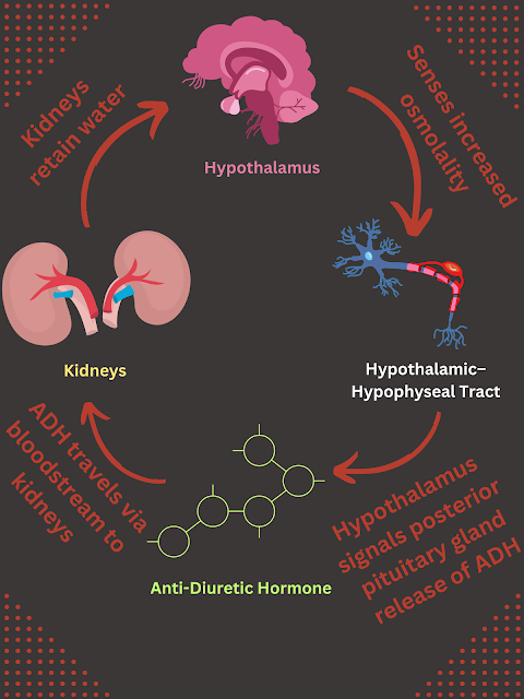
The steps of the release of ADH is written out below and followed by an infographic:

  1. The hypothalamus senses increased osmolarity of the bloodstream (increased concentration of solutes)
  2. The osmoreceptors in the hypothalamus shrink (due to a higher osmolarity in the bloodstream versus inside these cells)
  3. The hypothalamus sends an electric signal via the neurons to the poster pituitary gland to release ADH
  4. The ADH enters the bloodstream and arrives at the kidneys
  5. The kidneys increase the number of aquaporins in the distal tubules and collecting ducts
  6. More water is reabsorbed into the body and less water is urinated out


    Image of the above steps

 πŸŒŠ

Why is ADH Important?

ADH is extremely important because it allows the body to have the correct amount of water in the body.  Water is the basis of life and if there is not enough water in the body the body will not be able to survive very long.


As an aside, alcohol and caffeine are two substances that block the release of ADH from the posterior pituitary gland.  Due to this, the kidneys are not able to reabsorb water into the bloodstream which causes increased urination of very dilute urine.


Think about the last time you had an adult beverage or drank tea/coffee.  Did you have to pee shortly after?  Was your urine very light in color?  You can thank the blocking of the release of ADH for that change.  Cheers!


🍻🍡

Dr. Haus's Unsolicited Life Advice:  Drink water when drinking alcohol


What is Erythropoietin (EPO) and Why is it Important?

Picture of a kayak next to a lake

Photo by Dr. Haus

Erythropoietin (EPO)

Erythropoietin, called EPO for short, is a hormone that is released by the kidneys that tells the bone marrow to increase erythropoiesis.  Erythropoiesis is the production of new red blood cells and this occurs in the bone marrow of the body.


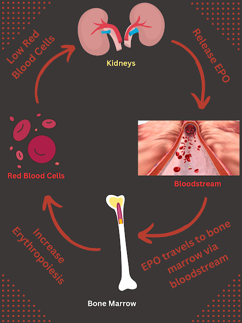
The steps of the release of EPO is written out below and followed by an infographic:

  1. The kidneys sense less oxygen arriving to the kidneys in the bloodstream
  2. The kidneys release EPO from the peritubular cells
  3. The EPO travels in the bloodstream to the bone marrow
  4. The EPO signals the bone marrow to increase the rate of erythropoiesis
  5. The bone marrow increase red blood cell synthesis
  6. Once the oxygen levels in the bloodstream increase the amount of oxygen arriving in the bloodstream to the kidneys goes back to normal and the peritubular cells stop releasing EPO


    Infographic of the steps of EPO release which are written out above

 πŸ©Έ

Why is Erythropoietin Important?

EPO is extremely important because it allows the body to have the correct amount of red blood cells.  The red blood cells are needed to bring oxygen to all the cells of the body to keep them alive.  Having too few (or too many) red blood cells is detrimental to the body.


As an aside, athletes can "dope" their blood with EPO (think Lance Armstrong here).  When athletes take EPO as a performance enhancement drug, they are increasing the number of red blood cells in their body which allows them to bring more oxygen to the cells of their body.  This allows these athletes to have more oxygen allowing them to fatigue less quickly.  This is obviously not legal and is cheating.


Now, you may also know that many athletes try to train at higher altitudes.  This is a strategic decision because at higher altitudes there is less oxygen in the air.  Due to this, people and animals that live at higher altitudes have more red blood cells in their body as their kidneys respond to having less oxygen in their system than a(n) person/animal at a lower altitude.  This is one way athletes strengthen their bodies for competition.  If you are working as a medical professional at a higher altitude it is important to know that the hematocrit (volume of red blood cells in the bloodstream) of patients at higher altitudes should be higher than at lower altitudes.


It is also unhealthy to have too many red blood cells in the bloodstream and this is called polycythemia.  This is dangerous because the blood becomes thicker, think sludge flowing through your body.  This puts a patient at risk for a blood clot, a fatal condition where blood flow is blocked causing no oxygen or nutrients from arriving to the body.

Dr. Haus's Unsolicited Life Advice:  Don’t do illegal drugs

What is the RAAS and Why is it Important?

Photo by Dr. Haus

Renin-Angiotensin-Aldosterone System (RAAS)

The Renin-Angiotensin-Aldosterone System, also called RAAS, is a hormone system that helps maintain blood pressure and blood volume.  RAAS is one of the most important regulatory systems of the body and I always joke that it is the only information you need to know to be a doctor (it is both a joke and not a joke...).


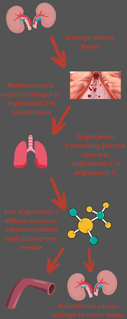
The steps of the RAAS are written out below and followed by an infographic:

  1. The kidneys release renin due to decreased blood flow to the kidneys, low sodium detected by the kidneys, and/or decreased blood pressure to the kidneys
  2. The renin converts angiotensinogen to angiotensin 1 in the bloodstream
  3. Angiotensin 1 arrives at the lungs where the Angiotensin Converting Enzyme (ACE) converts angiotensin 1 to angiotensin 2
  4. Angiotensin 2 has two effects: systemic vasoconstriction and the release of aldosterone from the kidneys
  5. Aldosterone increases sodium reabsorption and potassium secretion in the kidneys
  6. More water is reabsorbed into the body and less water is urinated out


    Picture of the above steps of RAAS

 πŸŒŠ

Why is RAAS Important?

The RAAS is extremely important for maintaining blood pressure in the body.  If blood pressure gets too low, the organs of the body will not be able to get all the blood flow they need for oxygen and nutrients.

πŸ«€

Dr. Hau's Unsolicited Life Advice:  Seriously, memorize RAAS

Thursday, October 26, 2023

Thoughtful Thursday: Florence Feels Feeble (Case 2)

Picture of stuffed alpaca named Dr. Pocky with stethoscope 

Photo of Professor Pocky taken by Dr. Haus

Click For Explanation of Case Flow


Click for Test Case Tuesday:  Florence Feels Feeble


Diagnostic Testing Results - Reminder: 


CBC (Complete Blood Count)

Red Blood Cells - Low (Anemia)


CHEM (Chemistry)

Renal values consistent with a renal (intrarenal) injury

Calcium - Low (hypocalcemia)


Urinalysis (UA)

Electrolyte values consistent with a renal (intrarenal) injury

Extremely dilute urine

πŸ§ͺ

Diagnosis:

✨CHRONIC KIDNEY DISEASE (FAILURE)✨


Pathophysiological Point:

Florence is suffering from a disease where her body's nephrons (kidney cells) are slowly dyingThis means her kidneys are slowly shutting down.


 Questions:

Level 1 Questions:

  1.  Explain what the kidneys do and why they are important.

  2.  If Florence has dilute urine, what does she need to have access to AT ALL TIMES?  (HINT:  Dilute urine means Florence is peeing out more water than normal)

  3.  Challenge question, why does Florence have a low number of red blood cells in her body?  (HINT:  For this one you will need to do some information gathering on the kidneys' different jobs)


Level 2 Questions:

  1.  Explain pathophysiologically why Florence has pale gums on physical exam and is anemic on blood work.  Be sure to explain all the steps of the process that is NOT occurring in Florence due to her chronic kidney disease.

  2.  Urea is a waste product that is normally urinated out of a patient's body via their kidneys and is an acid.  What acid-base status would you expect in Florence's bloodstream?

  3.  Challenge question, what TWO compensatory mechanisms are activated in Florence's body (there are two – assume the juxtaglomerular cells are still functional)?  (HINT:  Think about Florence's dilute urine)


Level 3 Questions:

  1.   Describe the treatment plan you would recommend for Florence and why you are recommending each part of your treatment plan.  Please answer this question as if you are speaking to a professional colleague.

  2.  Describe your recommended treatment plan and why you are recommending each part of your treatment plan.  Please answer this question as if you are explaining it to a client/patient without a scientific background.

  3.  Explain the pathophysiology of at least TWO (2) different etiologies of Chronic Kidney Disease.


Day 2 Conclusion

Before Conclusion Caturday think about your...

❓Answers to the above questions

πŸ“š Review Material Related to Florence's Case


Conclusion Caturday:  Florence Feels Feeble (link will go live Saturday, 10/28/2023)


Dr. Hau's Unsolicited Life Advice:  The only thing you need to know to be a doctor is the RAAS System

Tuesday, October 24, 2023

Test Case Tuesday: Florence Feels Feeble (Case 2)

Picture of stuffed Alpaca named Professor Pocky wearing a stethoscope
Picture of Professor Pocky taken by Dr. Haus

Click For Explanation of Case Flow


Signalment:

Florence is a 10-year-old female intact cheetah living in a sanctuary in Africa

🐱

History:

Over the past few months, the sanctuary workers have noticed that Florence is losing weight and showing signs of muscle atrophy (her muscles are wasting/thinning).  For the past week, Florence has been eating much less and she vomited twice.  Florence will sit at the side of her watering hole and drink a lot of water multiple times a day (she never did that before).  It is hard to say if Florence is urinating more as she is in an open living situation with multiple other cheetah friends.​

None of the other cheetahs in the habitat are showing any signs of disease.​

🌊 

Physical Exam Findings:


Attitude:

Lethargic (tired and blah) πŸ₯±


Body Condition Score (BCS) ☠️

3/9


Gum Color (Mucus Membrane Color):

Pale (light pink) 🩷


🩺


πŸ›‘STOP and brainstorm what diagnostic tests you would like to perform on Florence πŸ›‘


Caution Sign Saying What Diagnostic Testing Should Be Performed?


Diagnostic Testing Results:


CBC (Complete Blood Count)

Red Blood Cells - Low (Anemia)


CHEM (Chemistry)

Renal values consistent with a renal (intrarenal) injury

Calcium - Low (hypocalcemia)


Urinalysis (UA)

Electrolyte values consistent with a renal (intrarenal) injury

Extremely dilute urine 

πŸ§ͺ

Day 1 Conclusion

Before Thoughtful Thursday think about your...

πŸ“‹ Differentials List

πŸ§ͺ Further Diagnostic Testing

πŸ“š Review Material Related to Florence's Case (i.e. blood cell types, specific diseases of interest, categories of kidney disease, causes of increased drinking (polydipsia), anemia, personal interests, confusing points, etc.)


Thoughtful Thursday:  Florence Feels Feeble (link will go live Thursday, 10/26/2023)


Dr. Haus's Unsolicited Life Advice:  Know your renal physiology, it will always bite you in the booty

Conclusion Caturday: Vince Vomits Valiantly (Case 7)

Photo of TA Douglas (Left) and Professor Pocky (Right) taken by Dr. Haus   Click For Explanation of Case Flow   Click for Test Case Tuesday:...